Popular Posts

Popular Posts

Pages

Total Pageviews

Tuesday, February 28, 2012

Paper..How it is made..Wikipaedia.

paper mill is a factory devoted to making paper from vegetable fibres such as wood pulp, old rags and other ingredients using a Fourdrinier machine or other type of paper machine.
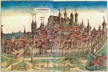


Stromer's paper mill, the building complex at the far right bottom, in the Nuremberg Chronicle of 1493. Due to their noise and smell, papermills were required by medieval law to be erected some distance from the city walls.
mid Nineteenth century paper mill, the Forest Fibre Company, in Berlin, New Hampshire.
Basement of paper mill in Sault Ste. Marie, Ontario. Pulp and paper manufacture involves a great deal of humidity, which presents a preventive maintenance andcorrosion challenge.
The Diamond Sutra of the Chinese Tang Dynasty, the oldest dated printed book in the world, found at Dunhuang, from 868 CE.
Papermaking is the process of making paper, a substance which is used universally today for writing and packaging.
In papermaking a dilute suspension of fibres in water is drained through a screen, so that a mat of randomly interwoven fibres is laid down. Water is removed from this mat of fibres by pressing and drying to make paper. Since the invention of the Fourdrinier machine in the 19th century, most paper has been made from wood pulp because of cost. But other fibre sources such as cotton and textiles are used for high-quality papers. One common measure of a paper's quality is its non-woodpulp content, e.g., 25% cotton, 50% rag, etc.
Papermaking, regardless of the scale on which it is done, involves making a dilute suspension of fibres in water and allowing this suspension to drain through a screen so that a mat of randomly interwoven fibres is laid down. Water is removed from this mat of fibres by pressing and drying to make paper.
An illustration from 105 AD depicting the papermaking process as designed by Cai Lun.
First the fibres are suspended in water to form a slurry in a large vat. The mold is a wire screen in a wooden frame (somewhat similar to an old window screen), which is used to scoop some of the slurry out of the vat. The slurry in the screen mold is sloshed around the mold until it forms a uniform thin coating. The fibres are allowed to settle and the water to drain. When the fibres have stabilized in place but are still damp, they are turned out onto a felt sheet which was generally made of an animal product such as wool or rabbit fur, and the screen mold immediately reused. Layers of paper and felt build up in a pile (called a 'post') then a weight is placed on top to press out excess water and keep the paper fibres flat and tight. The sheets are then removed from the post and hung or laid out to dry. A step-by-step procedure for making paper with readily available materials can be found online.[13]
When the paper pages are dry, they are frequently run between rollers (calendered) to produce a harder writing surface. Papers may be sized with gelatin or similar to bind the fibres into the sheet. Papers can be made with different surfaces depending on their intended purpose. Paper intended for printing or writing with ink is fairly hard, while paper to be used for water color, for instance, is heavily sized, and can be fairly soft.
The wooden frame is called a "deckle". The deckle leaves the edges of the paper slightly irregular and wavy, called "deckle edges", one of the indications that the paper was made by hand. Deckle-edged paper is occasionally mechanically imitated today to create the impression of old-fashioned luxury. The impressions in paper caused by the wires in the screen that run sideways are called "laid lines" and the impressions made, usually from top to bottom, by the wires holding the sideways wires together are called "chain lines". Watermarks are created by weaving a design into the wires in the mold. This is essentially true of Oriental molds made of other substances, such as bamboo. Hand-made paper generally folds and tears more evenly along the laid lines.
Hand-made paper is also prepared in laboratories to study papermaking and to check in paper mills the quality of the production process. The "handsheets" made according to TAPPI Standard T 205[14] are circular sheets 15.9 cm (6.25 in) in diameter and are tested on paper characteristics as paper brightness, strength, degree of sizing.[15]

Paper banknotes

Most banknotes are made from cotton paper (see also paper) with a weight of 80 to 90 grams per square meter. The cotton is sometimes mixed with linen,abaca, or other textile fibres. Generally, the paper used is different from ordinary paper: it is much more resilient, resists wear and tear (the average life of a banknote is two years),[16] and also does not contain the usual agents that make ordinary paper glow slightly under ultraviolet light. Unlike most printing and writing paper, banknote paper is infused with polyvinyl alcohol or gelatin to give it extra strength. Early Chinese banknotes were printed on paper made ofmulberry bark and this fiber is used in Japanese banknote paper today.
Most banknotes are made using the mould made process in which a watermark and thread is incorporated during the paper forming process. The thread is a simple looking security component found in most banknotes. It is however often rather complex in construction comprising fluorescent, magnetic, metallic and micro print elements. By combining it with watermarking technology the thread can be made to surface periodically on one side only. This is known as windowed thread and further increases the counterfeit resistance of the banknote paper. This process was invented by Portals, part of the De La Rue group in the UK. Other related methods include watermarking to reduce the number of corner folds by strengthening this part of the note, coatings to reduce the accumulation of dirt on the note, and plastic windows in the paper that make it very hard to copy.

2004..Nobel Prize for medicine and physiology..MRI


Paul C. Lauterbur
Sir Peter Mansfield

Paul C. Lauterbur

Sir Peter Mansfield

The Nobel Prize in Physiology or Medicine 2003 was awarded jointly to Paul C. Lauterbur and Sir Peter Mansfield "for their discoveries concerning magnetic resonance imaging"
      

Summary

Imaging of human internal organs with exact and non-invasive methods is very important for medical diagnosis, treatment and follow-up. This year's Nobel Laureates in Physiology or Medicine have made seminal discoveries concerning the use of magnetic resonance to visualize different structures. These discoveries have led to the development of modern magnetic resonance imaging, MRI, which represents a breakthrough in medical diagnostics and research.
Atomic nuclei in a strong magnetic field rotate with a frequency that is dependent on the strength of the magnetic field. Their energy can be increased if they absorb radio waves with the same frequency (resonance). When the atomic nuclei return to their previous energy level, radio waves are emitted. These discoveries were awarded the Nobel Prize in Physics in 1952. During the following decades, magnetic resonance was used mainly for studies of the chemical structure of substances. In the beginning of the 1970s, this year’s Nobel Laureates made pioneering contributions, which later led to the applications of magnetic resonance in medical imaging.
Paul Lauterbur (born 1929), Urbana, Illinois, USA, discovered the possibility to create a two-dimensional picture by introducing gradients in the magnetic field. By analysis of the characteristics of the emitted radio waves, he could determine their origin. This made it possible to build up two-dimensional pictures of structures that could not be visualized with other methods.
Peter Mansfield (born 1933), Nottingham, England, further developed the utilization of gradients in the magnetic field. He showed how the signals could be mathematically analysed, which made it possible to develop a useful imaging technique. Mansfield also showed how extremely fast imaging could be achievable. This became technically possible within medicine a decade later.
MRI
MRI is used for imaging of all organs in the body.
Magnetic resonance imaging, MRI, is now a routine method within medical diagnostics. Worldwide, more than 60 million investigations with MRI are performed each year, and the method is still in rapid development. MRI is often superior to other imaging techniques and has significantly improved diagnostics in many diseases. MRI has replaced several invasive modes of examination and thereby reduced the risk and discomfort for many patients.

Nuclei of hydrogen atoms

Water constitutes about two thirds of the human body weight, and this high water content explains why magnetic resonance imaging has become widely applicable to medicine. There are differences in water content among tissues and organs. In many diseases the pathological process results in changes of the water content, and this is reflected in the MR image.
Water is a molecule composed of hydrogen and oxygen atoms. The nuclei of the hydrogen atoms are able to act as microscopic compass needles. When the body is exposed to a strong magnetic field, the nuclei of the hydrogen atoms are directed into order – stand "at attention". When submitted to pulses of radio waves, the energy content of the nuclei changes. After the pulse, a resonance wave is emitted when the nuclei return to their previous state.
The small differences in the oscillations of the nuclei are detected. By advanced computer processing, it is possible to build up a three-dimensional image that reflects the chemical structure of the tissue, including differences in the water content and in movements of the water molecules. This results in a very detailed image of tissues and organs in the investigated area of the body. In this manner, pathological changes can be documented.

Rapid development within medicine

The medical use of magnetic resonance imaging has developed rapidly. The first MRI equipments in health were available at the beginning of the 1980s. In 2002, approximately 22 000 MRI cameras were in use worldwide, and more than 60 million MRI examinations were performed.
A great advantage with MRI is that it is harmless according to all present knowledge. The method does not use ionizing radiation, in contrast to ordinary X-ray (Nobel Prize in Physics in 1901) or computer tomography (Nobel Prize in Physiology or Medicine in 1979) examinations. However, patients with magnetic metal in the body or a pacemaker cannot be examined with MRI due to the strong magnetic field, and patients with claustrophobia may have difficulties undergoing MRI.

Especially valuable for examination of the brain and the spinal cord

Today, MRI is used to examine almost all organs of the body. The technique is especially valuable for detailed imaging of the brain and the spinal cord. Nearly all brain disorders lead to alterations in water content, which is reflected in the MRI picture. A difference in water content of less than a percent is enough to detect a pathological change.
In multiple sclerosis, examination with MRI is superior for diagnosis and follow-up of the disease. The symptoms associated with multiple sclerosis are caused by local inflammation in the brain and the spinal cord. With MRI, it is possible to see where in the nervous system the inflammation is localized, how intense it is, and also how it is influenced by treatment.
Imaging of the brain and the spinal cord
Examination with MRI is especially valuable for detailed imaging of the brain and the spinal cord.
Another example is prolonged lower back pain, leading to great suffering for the patient and to high costs for the society. It is important to be able to differentiate between muscle pain and pain caused by pressure on a nerve or the spinal cord. MRI examinations have been able to replace previous methods which were unpleasant for the patient. With MRI, it is possible to see if a disc herniation is pressing on a nerve and to determine if an operation is necessary.

Important preoperative tool

Since MRI yields detailed three-dimensional images, it is possible to get distinct information on where a lesion is localized. Such information is valuable before surgery. For instance, in certain microsurgical brain operations, the surgeon can operate with guidance from the MRI results. The images are detailed enough to allow placement of electrodes in central brain nuclei in order to treat severe pain or to treat movement disorders in Parkinson's disease.

Improved diagnostics in cancer

MRI examinations are very important in diagnosis, treatment and follow-up of cancer. The images can exactly reveal the limits of a tumour, which contributes to more precise surgery and radiation therapy. Before surgery, it is important to know whether the tumour has infiltrated the surrounding tissue. MRI can more exactly than other methods differentiate between tissues and thereby contribute to improved surgery.
MRI has also improved the possibilities to ascertain the stage of a tumour, and this is important for the choice of treatment. For example, MRI can determine how deep in the tissue a colon cancer has infiltrated and whether regional lymph nodes have been affected.

Reduced suffering for patients

MRI can replace previously used invasive examinations and thereby reduce the suffering for many patients. One example is investigation of the pancreatic and bile ducts with contrast media injection via an endoscope. This can in some cases lead to serious complications. Today, corresponding information can be obtained by MRI.
Diagnostic arthroscopy (examination with an optic instrument inserted into the joint) can be replaced by MRI. In the knee, it is possible to perform detailed MRI studies of the joint cartilage and the cruciate ligaments. Since no invasive instrument is needed in MRI, the risk of infection is eliminated.