Popular Posts

Popular Posts

Pages

Total Pageviews

Sunday, January 23, 2011

Eli Whitney..Invented the Cotton Gin...1793

  • The cotton gin is a machine that separates cotton fibers from seedpods and sometimes sticky seeds, a job previously done by hand. These seeds are either used again to grow more cotton or, if badly damaged, disposed. The cotton gin uses a combination of a wire screen and small wire hooks to pull the cotton through the screen, while brushes continuously remove the loose cotton lint to prevent jams. In 1793, Eli Whitney invented the cotton gin and later received a patent on March 14, 1794. Whitney's cotton gin could have possibly ignited a revolution in the cotton industry and the rise of "King Cotton" as the main cash crop in the South. However, it never made him rich. Instead of buying his machine, farmers built inferior versions of their own which led to the increasing need for African-American slave labor.

Liquid Crystal Display.(LCD); Invention

Liquid Asset: It all began with a carrot. In 1888, an Austrian botanist named Friedrich Reinitzer discovered liquid crystals (strictly speaking, not crystals at all, but liquids whose molecules scatter light the same way crystals do) in cholesterol he had extracted from carrots. But it took George Heilmeier and a group of fellow scientists at RCA’s David Sarnoff Research Center in Princeton to envision the use of those crystals in wristwatch and other displays, and to produce the world’s first LCD in 1968. Heilmeier went on to earn fifteen patents; help develop stealth aircraft, lasers, and artificial intelligence for the Department of Defense; and head New Jersey-based Bellcore (formerly Bell Communications Research). But he remains best known as the man who enabled us to spell hello on a calculator.

How Do Medical Image Machines Work?

How Do Medical Image Machines Work?

Radiographs, utilizing X-rays, are often used to view the heart and lungs.
an xray of a chest image by alma_sacra from Fotolia.com
The ability to perceive the state of structures within the human body without opening it surgically has transformed medicine's ability to diagnose disease. While the practice of medicine has existed in some form for thousands of years, medical imaging has been around only since 1895. Starting with radiography, medical imaging has branched into several modalities, each best suited for various conditions. Kinds of imaging machines share similarities and differences in the way they work.

    Physics

  1. Generally, most medical imaging functions by permeating the body with energy that is either absorbed, transmitted through or reflected back. The subtle changes in this energy is detected by sensitive chemicals, electronic sensors or other detectors and sometimes analyzed by computers. The type of energy used varies according to the type of pathology suspected, area to be examined or other factors.
  2. X-Rays

  3. The use of X-rays in medical examination dates to 1895. Dr. Wilhelm Conrad Rontgen (a physicist, not a physician) was experimenting with vacuum tubes that emitted radiation when he discovered that some of the radiation was not only invisible but could penetrate cloth, wood, paper and even flesh. As he continued to experiment with the equipment, he found that the rays could be directed at an object (or person) to be examined and would produce a shadow that could be captured on film. Where the rays encounter little resistance, they leave a dark area. Thin tissue, air, lungs and other such structures appear dark. Denser tissue, such as heart muscle, visceral organs and bones, absorb the energy and appear on developed film as light areas.
  4. Computerized Tomography

  5. Computerized tomography, also called CT or CAT scan, works by employing the same kind of energy. X-ray frequency radiation is essentially the same kind of radiation as visible light but is at a much more energetic frequency, allowing it to penetrate objects opaque to light. In computerized tomography, X-ray frequency radiation directed through the body is detected by sensors. Both the emitters and detectors rotate around the body of the subject, and this motion produces data about the structures from a variety of perspectives. No film is produced as in the simpler radiograph (X-ray film). Instead, computers use sophisticated algorithms to produce a mathematical model of the patient, which is displayed on computer screens or printed for easier viewing or transport.
  6. Sonography

  7. The waves of energy capable of penetrating the body don't have to be electromagnetic in nature. Sonography, also known as ultrasound, uses sound waves, which are easily transmitted by fluid and, because the human body is composed mostly of water, most parts of the anatomy. Soft tissue is most easily imaged by sonography, which also has the advantage of being far more portable than computerized tomography or X-ray. Though early work into sonography was done in the 1940s, a practical scanner for producing images was not fielded until the 1960s.
  8. Magnetic Resonance Imaging

  9. Magnetic resonance imaging (MRI) is sometimes regarded as the most esoteric of the modalities in common use. The specifics of MRI are quite complicated; in short, though, the subject is placed in a very strong magnetic field and then exposed to radio-frequency radiation. This process causes the hydrogen atoms in the body to emit radiation. The sensors in the machine detect the location, quantity and frequency of these emissions, and a powerful computer uses this raw data to construct a virtual model of the subject's body. MRI is effective for viewing both soft and bony tissue. Though the word "radiation" is used, it's important to note that ordinary light and radio waves are radiation, and they are harmless. Like them, the radiation used by MRI has not been shown to cause side effects or illness, and it is a major advantage of the technique.

BERNERS-LEE, TIM inventor of the WWW

BERNERS-LEE, TIM
Tim Berners-Lee (1955, London, England - ) invented the World Wide Web. His first version of the Web was a program named "Enquire," short for "Enquire Within Upon Everything". At the time, Berners-Lee was working at CERN, the European Particle Physics Laboratory located in Geneva, Switzerland. He invented the system as a way of sharing scientific data (and other information) around the world, using the Internet, a world-wide network of computers, and hypertext documents. He wrote the language HTML (HyperText Mark-up Language), the basic language for the Web, and devised URL's (universal resource locators) to designate the location of each web page. HTTP (HyperText Transfer Protocol) was his set of rules for linking to pages on the Web. After he wrote the first browser in 1990, the World Wide Web was up and going. Its growth was (and still is) phenomenal, and has changed the world, making information more accessible than ever before in history. Berners-Lee is now a Principal Research Scientist at the Laboratory for Computer Science at the MIT (Massachusetts Institute of Technology, in Cambridge, Massachusett, USA) and the Director of the W3 Consortium.

APGAR Scale for New Borns

APGAR SCALE
The Apgar scale is a standardized scale that is used to determine the physical status of an infant at birth. This simple, easy-to-perform test was devised in 1953 by Dr. Virginia Apgar (1909-1974), a professor of anesthesia at the New York Columbia-Presbyterian Medical Center. The Apgar scale is administered to a newborn at one minute after birth and five minutes after birth. It scores the baby's heart rate, respiration, muscle tone, reflex response, and color. This test quickly alerts medical personnel that the newborn needs assistance. APGAR, VIRGINIA
Dr. Virginia Apgar (1909-1974), a professor of anesthesiology at the New York Columbia-Presbyterian Medical Center, devised the Apgar Scale in 1953. The Apgar scale is a simple, easy-to-perform, standardized scale that is used to determine the physical status of an infant at birth. The Apgar scale is administered to a newborn at one minute after birth and five minutes after birth. It scores the baby's heart rate, respiration, muscle tone, reflex response, and color. This test quickly alerts medical personnel that the newborn baby needs assistance.

Locks.....by Mara Bishop, '00 and Amanda Payne Burton, '97

Tumbler Lock, Egypt, Iraq, Greece, 1000 BCE


This simple tumbler lock was probably invented around 1000 BCE, although a date of 2000 BCE has also been proposed. Many sources believe this lock was invented in Egypt, though locks of this type have been found in ruins in Iraq that predate those found in Egypt. After its invention and its movement into Egypt, the lock made its way into Greece. Here this new system was a great improvement over the previous locks, which were simply a board drawn across a door. From Greece the lock moved into Europe.

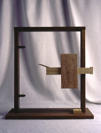
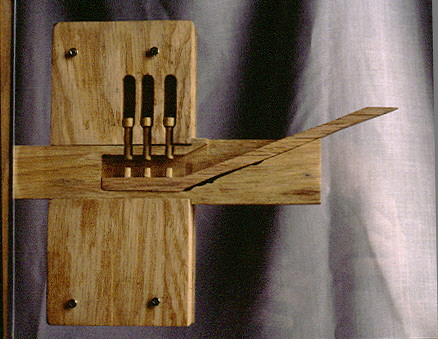
The lock is the first mechanical fastening for doors, and is said to be the only major European architectural improvement in classical times.The mechanism consists of a key and a lock. The key is simply a bit of wood with small pins, usually of brass. These pins enter small holes in the bolt and lift similar pins in the lock. The pins of the key push the lock pins out of the holes, and the bolt can be moved aside and the door opened. When leaving the building, the bolt is slid across the door jamb, and the pins fall into the grooves in the bolt. To unlock, the key is slid into the opening in the bolt and lifted up, which moves the bolt-pins out of the way.